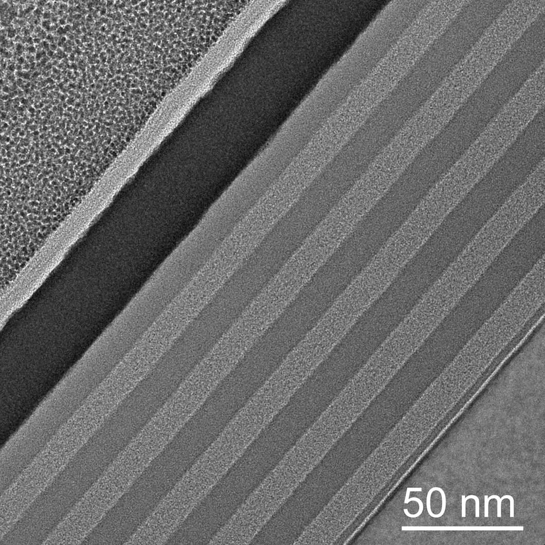
Cross-sectional TEM analysis
Cross-sectional transmission electron microscopy (cross-section TEM) is a method for obtaining high-resolution images of the cross-sections of larger materials. The area of interest is selected from a larger sample, prepared using a technique like FIB, and imaged with TEM. The method is widely used in materials science and nanotechnology.

Some of our TEM services
HR-TEM imaging
STEM-EDX
Particle size distribution with TEM
Negative staining TEM or cryo-TEM for liposomal powders
Prices excluding VAT.
What is cross-sectional TEM used for?
A transmission electron microscope (TEM) uses a high-energy electron beam to form high-resolution images of samples that are less than 100 nm thick. Preparation procedures for such thin samples can be complex, and sometimes the desired area for imaging is inside the sample, for example, when the boundary between layers in thin film materials is studied. In such cases, a cross-section of the sample needs to be prepared before imaging.
Cross-sectional TEM is widely used in material science, cell biology, and nanotechnology to image the internal structures of materials that are otherwise too large for TEM. One common application is determining the thickness and uniformity of thin films.
Cross-section preparation methods
There are two main methods to obtain a cross-sectional sample for TEM: microtomy and ion beam cutting.
A microtome machine uses blades to slice the sample into 60-100 nm thick chips. The blades are made of glass, steel, or diamond. Microtomes can be used to prepare samples from bones, minerals, and polymeric materials such as plastics. Sometimes, samples are molded in epoxy before microtomy.
A focused ion beam (FIB) is a nano-machining technique that uses a high-energy ion beam to mill the cross-sectional sample out from a larger object. A dual ion beam is used to carve a piece of the original sample. This piece is then removed with a carrier and mounted for thinning with the ion beam to achieve the required thickness.
Need cross-sectional TEM analyses?
Measurlabs offers high-quality testing services with TEM, cross-sectional TEM, and several additional electron microscopy techniques. Short turnaround times keep projects on schedule, while a broad range of analytical methods across industries allows all required analyses to be arranged under a single purchase order. Use the form below to start the discussion with our experts.
Suitable sample matrices
- Materials that are too large for normal TEM.
- Thin films
- Semiconductors
- Metals
- Bone
- Tissues
Ideal uses of cross-sectional TEM
- Cross sectional imaging of semiconductors.
- Imaging of thin film layers.
- High resolution imaging for failure analysis.
- Nanoparticle characterisation.
- Semiconductor research.
- Fault finding in material studies.
- Multilayer thin material analyses.
- Cell imaging in bone research.
Ask for an offer
Fill in the form, and we'll reply in one business day.
Have questions or need help? Email us at info@measurlabs.com or call our sales team.
Frequently asked questions
Cross-sectional TEM is used when the area of interest is inside of the sample, or the sample is too large for TEM imaging. TEM produces high-resolution 2D images.
Some materials may be too brittle for the preparation of the cross-sections. Cross-sectioning is also inherently destructive, and should therefore only be performed after relevant non-destructive test methods, such as micro-CT, have been employed.
A very wide range of materials can be examined with cross-sectional TEM. High-resolution images can be produced from rocks and minerals, bones, metals, thin films, and semiconductor materials.
Measurlabs offers a variety of laboratory analyses for product developers and quality managers. We perform some of the analyses in our own lab, but mostly we outsource them to carefully selected partner laboratories. This way we can send each sample to the lab that is best suited for the purpose, and offer high-quality analyses with more than a thousand different methods to our clients.
When you contact us through our contact form or by email, one of our specialists will take ownership of your case and answer your query. You get an offer with all the necessary details about the analysis, and can send your samples to the indicated address. We will then take care of sending your samples to the correct laboratories and write a clear report on the results for you.
Samples are usually delivered to our laboratory via courier. Contact us for further details before sending samples.