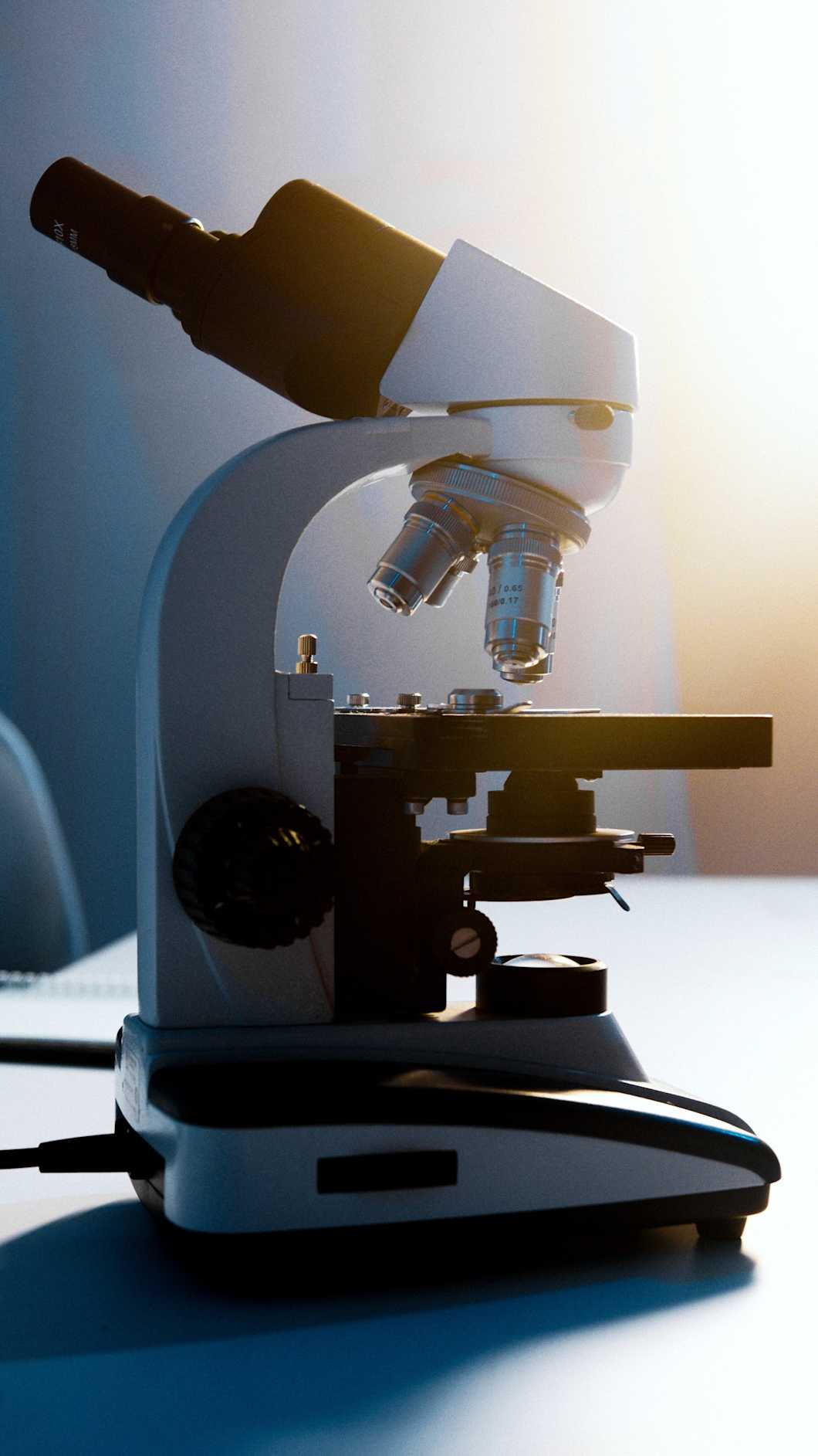
Optical microscopy
An optical microscope, also called a light microscope, uses visible light and lenses to create a magnified image of small objects that could not otherwise be seen with the naked eye. The magnification range of optical microscopy extends from 10x to 100x, which means that details in the 0.2-micrometer size range can be observed.

Optical microscopy is typically used in biological research and material science. It is often the first step to successful material analysis, giving a good overview of the material's microstructure and structure-property relationships.
The magnification range of the optical microscope is from 10x to 100x. This means that objects as small as 0.2 micrometers (0.2 thousands of a millimeter or 2 x 10⁻⁷ m) can be seen. For example, cells and large bacteria can be observed.
Optical microscopy is often used alongside other microscopy techniques with a higher resolution. For example, TEM and SEM use electrons to form images from much smaller objects, and optical microscopy is used to locate the area of interest from the sample. An optical microscope may also be combined with infrared spectroscopy for FTIR microscopy, which enables visualizing the chemical composition of a sample as a 2D map.
How does optical microscopy work?
In optical microscopy, visible light is transmitted through or reflected from the sample. The light then goes through a single lens or a series of lenses, which leads to a magnified view of the sample. The resulting image can be seen directly by the naked eye, or it can be imaged. Modern optical microscopes use digital imagining.
Sample preparation
Since biological structures are often colorless, they are typically stained with selective dyes to obtain better contrast for the image. The sample is also fixed, which means attaching it to a slide.
If the sample is transparent, a transmission mode is used. The specimen is usually a ten-micrometer thick slice. Absorption of light differs depending on the different regions and this arises in contrast. The method is used for the examination of rocks, polymers, and tissue samples.
Polarized light microscopy is a form of transmission mode. Contrast arises from differences in birefringence and thickness of the specimen. The method is used to observe grains, grain orientation, and thickness.
Reflected light microscopy is used for materials such as metals, ceramics, and composites. Variations in the surface arise in contrast when the light is reflected from the surface.
Limitations of optical microscopy
Optical microscopy has some drawbacks. The technique can produce images from materials that have enough contrast or from strongly refracting objects. Sample thickness is also restricted in the case of transmission mode to tens of micrometers. The resolution of the optical microscope is limited to 0.2 micrometers and the practical magnification limit is 1000x. For smaller objects, SEM, TEM, and AFM are preferred methods.
If you need microscopy services, do not hesitate to contact our experts using the form below. In addition to optical microscopy, we offer a wide range of electron microscopy analyses, as well as hot-stage microscopy for observing changes that occur with increasing temperature.
Suitable sample matrices
- Circuit boards
- Protective coatings and paints
- Cell and tissue samples
- Pollen
- Fibers such as asbestos
- Minerals and rock
Ideal uses of optical microscopy
- Initial surface analysis in materials research
- Structural studies in material science
- Examination of mineral and rock samples
- Study of cellular structures
- Observation of large bacteria
Ask for an offer
Fill in the form, and we'll reply in one business day.
Have questions or need help? Email us at info@measurlabs.com or call our sales team.
Frequently asked questions
Optical microscopy is used in material science to analyze structures. It can be used alone or in combination with other microscopy techniques, such as TEM or SEM. With an FTIR microscope, data can also be gathered on the chemical makeup of the sample.
Microscopy is also used for cell structure imaging. For example, it is a common tool in biology and medical diagnostics.
The resolution of the optical microscope is limited to 0.2 micrometers and the magnification is limited to 1000x.
Almost all kinds of samples are suitable for optical microscopy, as long as light passes through or reflects from the sample.
Measurlabs offers a variety of laboratory analyses for product developers and quality managers. We perform some of the analyses in our own lab, but mostly we outsource them to carefully selected partner laboratories. This way we can send each sample to the lab that is best suited for the purpose, and offer high-quality analyses with more than a thousand different methods to our clients.
When you contact us through our contact form or by email, one of our specialists will take ownership of your case and answer your query. You get an offer with all the necessary details about the analysis, and can send your samples to the indicated address. We will then take care of sending your samples to the correct laboratories and write a clear report on the results for you.
Samples are usually delivered to our laboratory via courier. Contact us for further details before sending samples.